Fundus Photo Of Diabetic Retinopathy: What It Can Show

Fundus photography involves taking a photo of the back of the eye to detect signs of diabetic retinopathy. It is also possible to capture an image of the retina using a smartphone.
Diabetic retinopathy is an eye disease that can develop in people with diabetes. Without treatment, it can lead to vision loss and blindness.
Fundus photography forms part of fundoscopy, an eye test that examines the back of the eye to check for signs of eye disease.
This article explains what fundus photos may show with diabetic retinopathy, examples, outlook, and when to contact a doctor.
Fundus photography creates an image of the internal structures of the eye, including the retina.
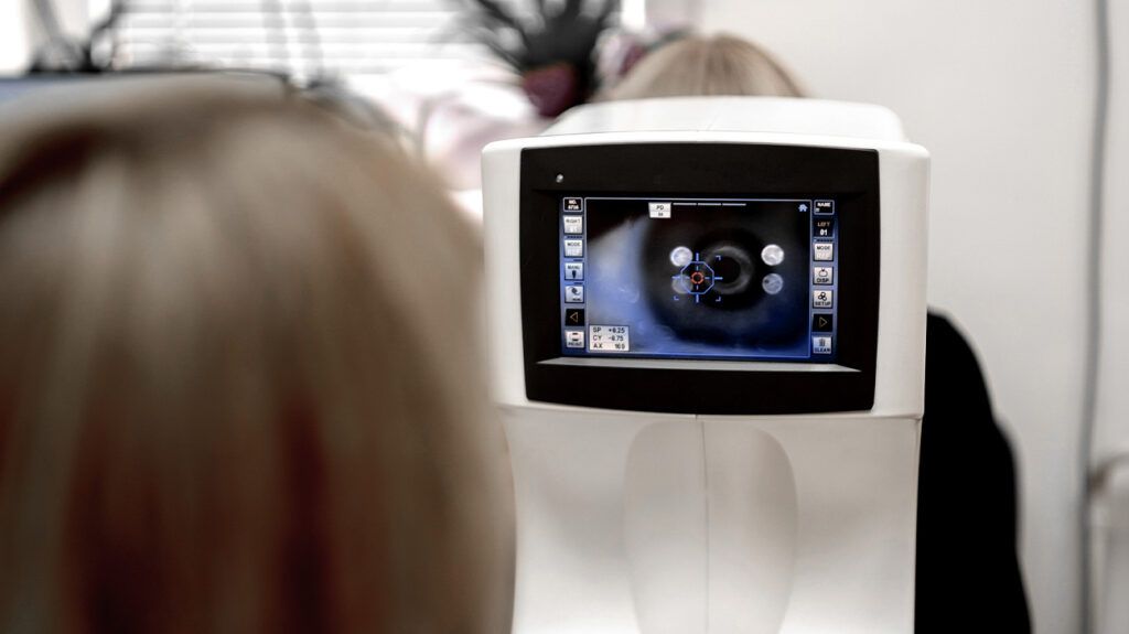
Fundoscopy is an eye test that examines the fundus, the inner surface at the back of the eye. During fundoscopy, an eye doctor uses a specialist fundus camera to take a picture of the back of the eye.
Fundus photography helps screen for, detect, and treat various eye conditions, including:
- diabetic retinopathy
- age-related macular degeneration
- glaucoma
Signs of diabetic retinopathy in fundus photos include:
- Microaneurysms: Microaneurysms are tiny lesions that appear as small, round, red dots with a clearly defined, regular margin. Microaneurysms are the earliest clinical lesions of diabetic retinopathy that doctors can detect.
- Hemorrhages: Diabetic retinopathy can damage blood vessels in the eye, weakening capillary walls, which can rupture. This causes retinal hemorrhage, which is bleeding within the retina. These hemorrhages may be flame shaped or dot-and-blot hemorrhages, which are dark red with sharp outlines.
- Hard exudates: Hard exudates are deposits of lipoproteins and lipids that have leaked from blood vessels. They are yellow, shiny, or waxy and may form a ring around a microaneurysm.
- Cotton wool spots: Cotton wool spots appear as white, fluffy patches on the retina. They can indicate a lack of blood supply to the area.
- Intraretinal microvascular abnormalities (IRMA): IRMA are abnormalities in the blood vessels in the retina, including abnormal growth or stretching of blood vessels.
- Neovascularization: This is the abnormal growth of new blood vessels in the eye. It is a sign of proliferative diabetic retinopathy, a more advanced stage of the disease.
Selfie fundus imaging (SFI) allows people to take a picture, or selfie, of the fundus using their smartphone.
With SFI, people apply special eye drops to dilate the pupil, then use a handheld smartphone to take a photo of the retina.
A 2022 study compared SFI with traditional fundus photography in 60 people with diabetes.
Researchers compared three techniques of fundus photography:
- handheld smartphone photos, which participants took themselves after a short video training session
- smartphone photos, which a trained technician took
- photos from a conventional digital fundus camera, which is the gold standard for fundus photography
Researchers found that the SFI photos, which participants easily took themselves, were similar to the photos a trained technician took.
SFI may make diabetic retinopathy screening more accessible and affordable.
Fundus photography may only take a few minutes, but the complete eye test takes longer. To perform fundoscopy, an eye doctor uses eye drops to dilate the eyes. It can take 20 to 30 minutes for the pupils to dilate fully.
Fundus photography requires dilating eye drops to work, which can temporarily cause blurry vision. This can make it unsafe to drive after a fundus test.
The side effects of the eye drops may last several hours. People should wait until their vision returns to normal before driving.
The fundus test is painless. People may have blurred vision and increased sensitivity to light for a short time after the test.
Anyone with diabetes is at risk of developing diabetic retinopathy. Getting dilated eye exams at least once yearly is important for detecting and treating diabetic retinopathy early.
If people with diabetes become pregnant or develop gestational diabetes, it is important to get a dilated eye exam as soon as possible. People can talk with a doctor to check whether they need extra eye tests during their pregnancy.
Diabetic retinopathy may not cause any symptoms in its early stages. As it progresses, people may experience symptoms such as:
- changes in vision, which may come and go, such as difficulty reading or seeing objects in the distance
- floaters, which may look like dark spots or cobweb-like strands
If people have any of these symptoms, they should contact a doctor as soon as possible.
Fundus photography is an important tool for diagnosing and treating diabetic retinopathy. Regular screening and prompt treatment can prevent 90% of blindness from diabetic retinopathy.
Managing diabetes through physical activity, eating a balanced diet, and taking any medication is also important in helping prevent or delay vision loss.
Fundus photography takes an image of the retina to check for signs of diabetic retinopathy.
Fundus photos can show signs of diabetic retinopathy, including spots of bleeding, areas of poor blood supply, and abnormalities in the blood vessels.
Getting regular dilated eye exams with diabetes is important for detecting and treating diabetic retinopathy early to prevent complications.


